Artlabeling Activity Oblique and Rectus Muscles of the Abdominal Wall Transverse Section
The Muscular Arrangement
Axial Muscles of the Abdominal Wall, and Thorax
Learning Objectives
Past the cease of this section, you will be able to:
- Identify the intrinsic skeletal muscles of the back and neck, and the skeletal muscles of the intestinal wall and thorax
- Identify the movement and function of the intrinsic skeletal muscles of the dorsum and neck, and the skeletal muscles of the abdominal wall and thorax
It is a circuitous job to balance the torso on two feet and walk upright. The muscles of the vertebral column, thorax, and abdominal wall extend, flex, and stabilize different parts of the body's trunk. The deep muscles of the core of the torso help maintain posture also as conduct out other functions. The brain sends out electrical impulses to these diverse muscle groups to control posture by alternate contraction and relaxation. This is necessary so that no single muscle group becomes drawn too quickly. If whatever one grouping fails to function, trunk posture will be compromised.
Muscles of the Abdomen
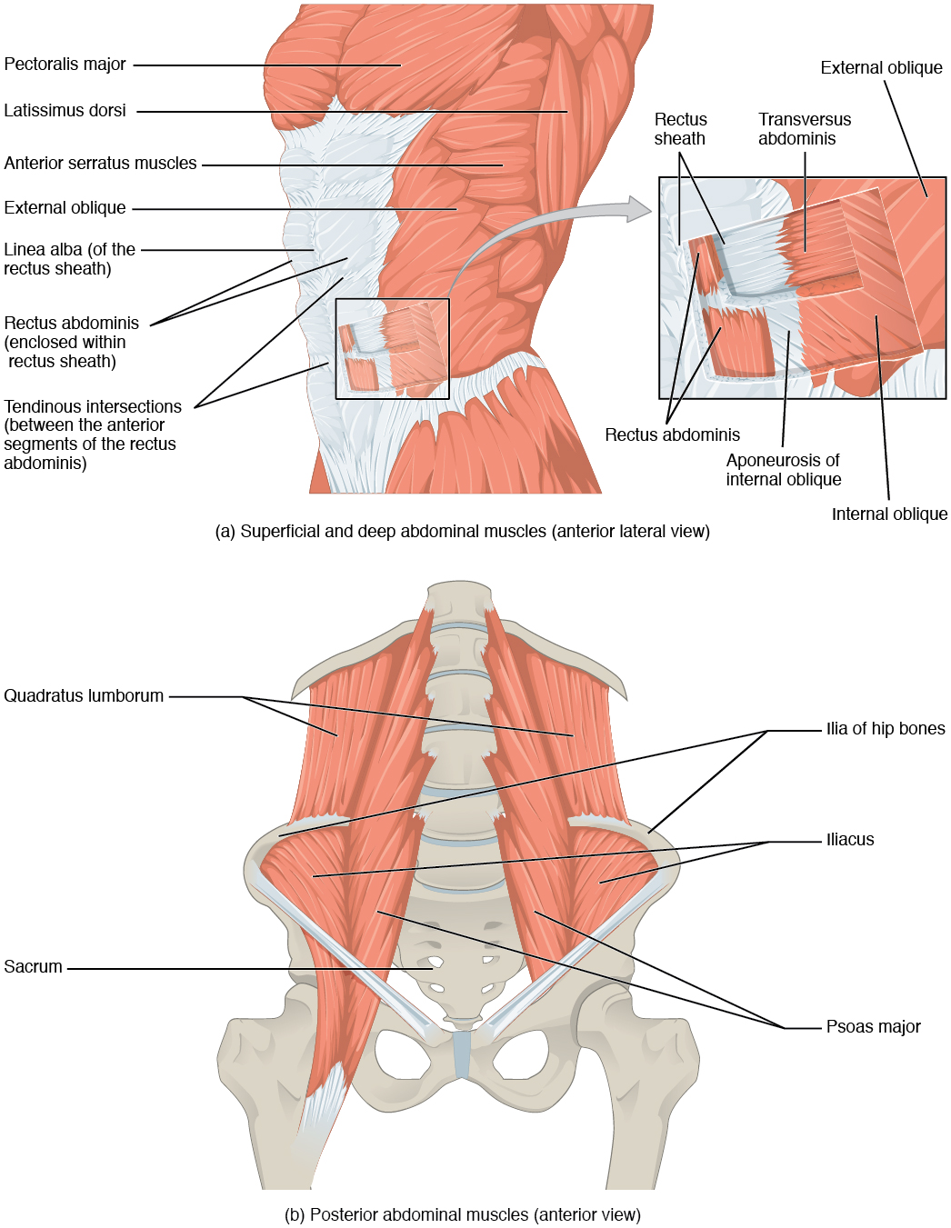
At that place are four pairs of intestinal muscles that cover the anterior and lateral abdominal region and meet at the anterior midline. These muscles of the anterolateral abdominal wall tin be divided into four groups: the external obliques, the internal obliques, the transversus abdominis, and the rectus abdominis ((Figure) and (Figure)).
Muscles of the Abdomen
(a) The anterior abdominal muscles include the medially located rectus abdominis, which is covered by a sheet of connective tissue called the rectus sheath. On the flanks of the torso, medial to the rectus abdominis, the intestinal wall is equanimous of three layers. The external oblique muscles form the superficial layer, while the internal oblique muscles course the middle layer, and the transverses abdominus forms the deepest layer. (b) The muscles of the lower back movement the lumbar spine but besides assist in femur movements.
| Muscles of the Belly | |||||
|---|---|---|---|---|---|
| Movement | Target | Target motion direction | Prime mover | Origin | Insertion |
| Twisting at waist; also bending to the side | Vertebral column | Supination; lateral flexion | External obliques; internal obliques | Ribs five–12; ilium | Ribs vii–10; linea alba; ilium |
| Squeezing abdomen during forceful exhalations, defecation, urination, and childbirth | Abdominal cavity | Compression | Transversus abdominus | Ilium; ribs 5–ten | Sternum; linea alba; pubis |
| Sitting upwardly | Vertebral column | Flexion | Rectus abdominis | Pubis | Sternum; ribs v and 7 |
| Angle to the side | Vertebral cavalcade | Lateral flexion | Quadratus lumborum | Ilium; ribs v–10 | Rib 12; vertebrae L1–L4 |
At that place are three apartment skeletal muscles in the antero-lateral wall of the belly. The external oblique, closest to the surface, extend inferiorly and medially, in the direction of sliding one's four fingers into pants pockets. Perpendicular to it is the intermediate internal oblique, extending superiorly and medially, the direction the thumbs usually become when the other fingers are in the pants pocket. The deep musculus, the transversus abdominis, is arranged transversely effectually the belly, like to the front of a belt on a pair of pants. This arrangement of 3 bands of muscles in different orientations allows various movements and rotations of the trunk. The three layers of musculus also help to protect the internal abdominal organs in an surface area where there is no os.
The linea alba is a white, fibrous band that is made of the bilateral rectus sheaths that join at the anterior midline of the body. These enclose the rectus abdominis muscles (a pair of long, linear muscles, unremarkably called the "sit-up" muscles) that originate at the pubic crest and symphysis, and extend the length of the torso's trunk. Each muscle is segmented by iii transverse bands of collagen fibers chosen the tendinous intersections. This results in the look of "six-pack abs," as each segment hypertrophies on individuals at the gym who do many sit-ups.
The posterior abdominal wall is formed by the lumbar vertebrae, parts of the ilia of the hip basic, psoas major and iliacus muscles, and quadratus lumborum muscle. This part of the cadre plays a key role in stabilizing the rest of the body and maintaining posture.
Career Connections
Concrete Therapists Those who have a muscle or joint injury will well-nigh likely be sent to a physical therapist (PT) afterwards seeing their regular medico. PTs have a master'due south degree or doctorate, and are highly trained experts in the mechanics of trunk movements. Many PTs also specialize in sports injuries.
If you injured your shoulder while yous were kayaking, the showtime matter a physical therapist would do during your first visit is to assess the functionality of the joint. The range of motility of a particular joint refers to the normal movements the joint performs. The PT will ask yous to abduct and adduct, revolve, and flex and extend the arm. The PT will note the shoulder'due south degree of function, and based on the assessment of the injury, volition create an appropriate physical therapy program.
The offset stride in physical therapy will probably be applying a heat pack to the injured site, which acts much like a warm-upwards to draw blood to the area, to enhance healing. You will be instructed to do a series of exercises to continue the therapy at dwelling, followed by icing, to decrease inflammation and swelling, which will continue for several weeks. When physical therapy is consummate, the PT will do an exit examination and ship a detailed report on the improved range of motion and render of normal limb function to your doctor. Gradually, as the injury heals, the shoulder will begin to function correctly. A PT works closely with patients to help them become back to their normal level of physical activity.
Muscles of the Thorax
The muscles of the chest serve to facilitate breathing past changing the size of the thoracic cavity ((Figure)). When you inhale, your chest rises because the cavity expands. Alternately, when yous exhale, your chest falls because the thoracic cavity decreases in size.
| Muscles of the Thorax | |||||
|---|---|---|---|---|---|
| Movement | Target | Target motility direction | Prime mover | Origin | Insertion |
| Inhalation; exhalation | Thoracic crenel | Compression; expansion | Diaphragm | Sternum; ribs 6–12; lumbar vertebrae | Central tendon |
| Inhalation;exhalation | Ribs | Elevation (expands thoracic cavity) | External intercostals | Rib superior to each intercostal muscle | Rib inferior to each intercostal muscle |
| Forced exhalation | Ribs | Move along superior/inferior axis to bring ribs closer together | Internal intercostals | Rib inferior to each intercostal muscle | Rib superior to each intercostal muscle |
The Diaphragm
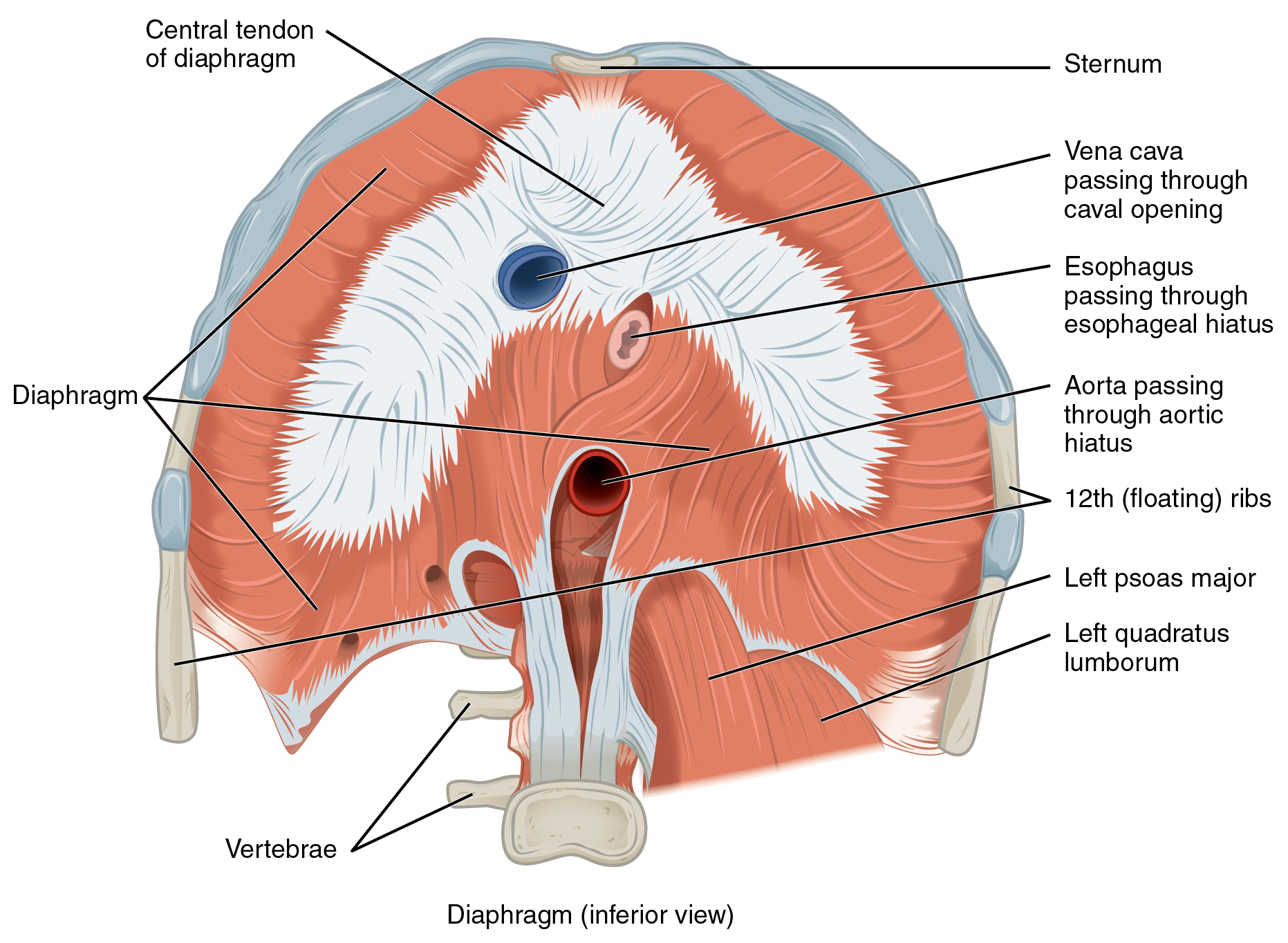
The change in book of the thoracic cavity during animate is due to the alternate contraction and relaxation of the diaphragm ((Effigy)). It separates the thoracic and abdominal cavities, and is dome-shaped at rest. The superior surface of the diaphragm is convex, creating the elevated floor of the thoracic cavity. The junior surface is concave, creating the curved roof of the intestinal crenel.
Muscles of the Diaphragm
The diaphragm separates the thoracic and abdominal cavities.
Defecating, urination, and even childbirth involve cooperation between the diaphragm and abdominal muscles (this cooperation is referred to as the "Valsalva maneuver"). You lot agree your breath by a steady contraction of the diaphragm; this stabilizes the volume and force per unit area of the peritoneal crenel. When the abdominal muscles contract, the pressure cannot push the diaphragm up, so information technology increases pressure on the intestinal tract (defecation), urinary tract (urination), or reproductive tract (childbirth).
The inferior surface of the pericardial sac and the inferior surfaces of the pleural membranes (parietal pleura) fuse onto the central tendon of the diaphragm. To the sides of the tendon are the skeletal musculus portions of the diaphragm, which insert into the tendon while having a number of origins including the xiphoid process of the sternum anteriorly, the inferior half-dozen ribs and their cartilages laterally, and the lumbar vertebrae and 12th ribs posteriorly.
The diaphragm too includes 3 openings for the passage of structures between the thorax and the belly. The inferior vena cava passes through the caval opening, and the esophagus and attached nerves pass through the esophageal hiatus. The aorta, thoracic duct, and azygous vein pass through the aortic hiatus of the posterior diaphragm.
The Intercostal Muscles
There are three sets of muscles, called intercostal muscles, which span each of the intercostal spaces. The primary role of the intercostal muscles is to assist in breathing by irresolute the dimensions of the rib cage ((Figure)).
Intercostal Muscles
The external intercostals are located laterally on the sides of the body. The internal intercostals are located medially near the sternum. The innermost intercostals are located deep to both the internal and external intercostals.
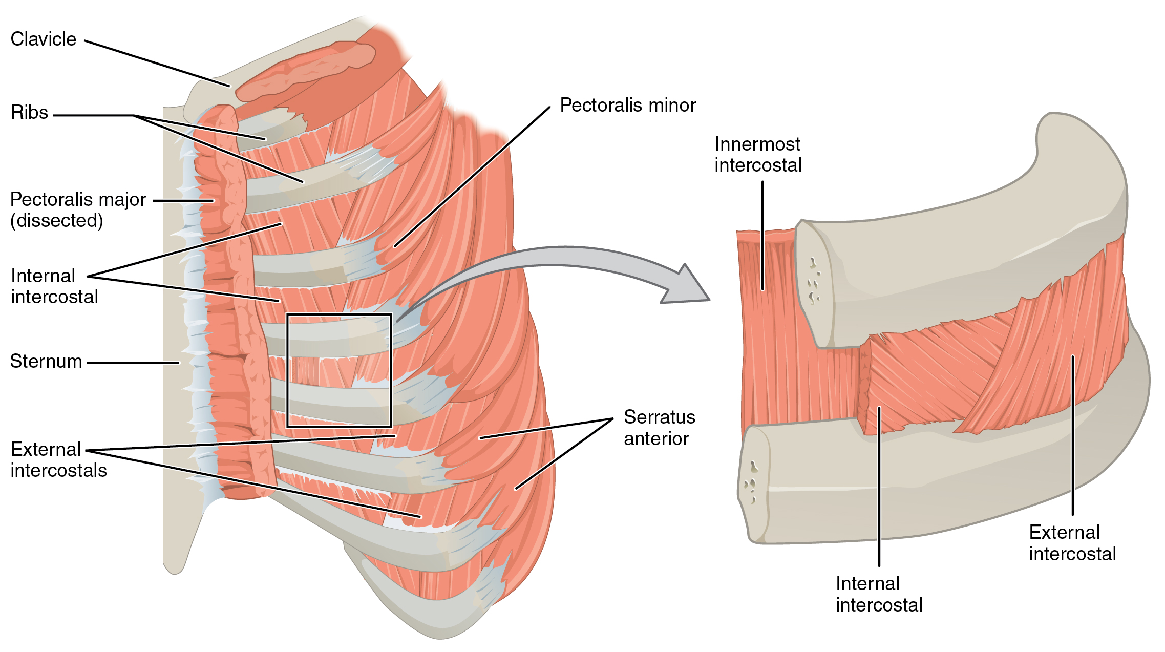
The 11 pairs of superficial external intercostal muscles aid in inspiration of air during animate because when they contract, they raise the rib cage, which expands it. The 11 pairs of internal intercostal muscles, but under the externals, are used for expiration because they describe the ribs together to constrict the rib cage. The innermost intercostal muscles are the deepest, and they deed as synergists for the action of the internal intercostals.
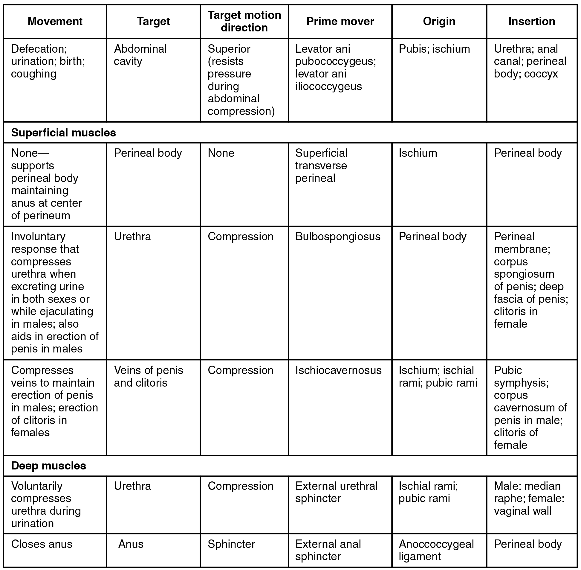
Muscles of the Pelvic Floor and Perineum
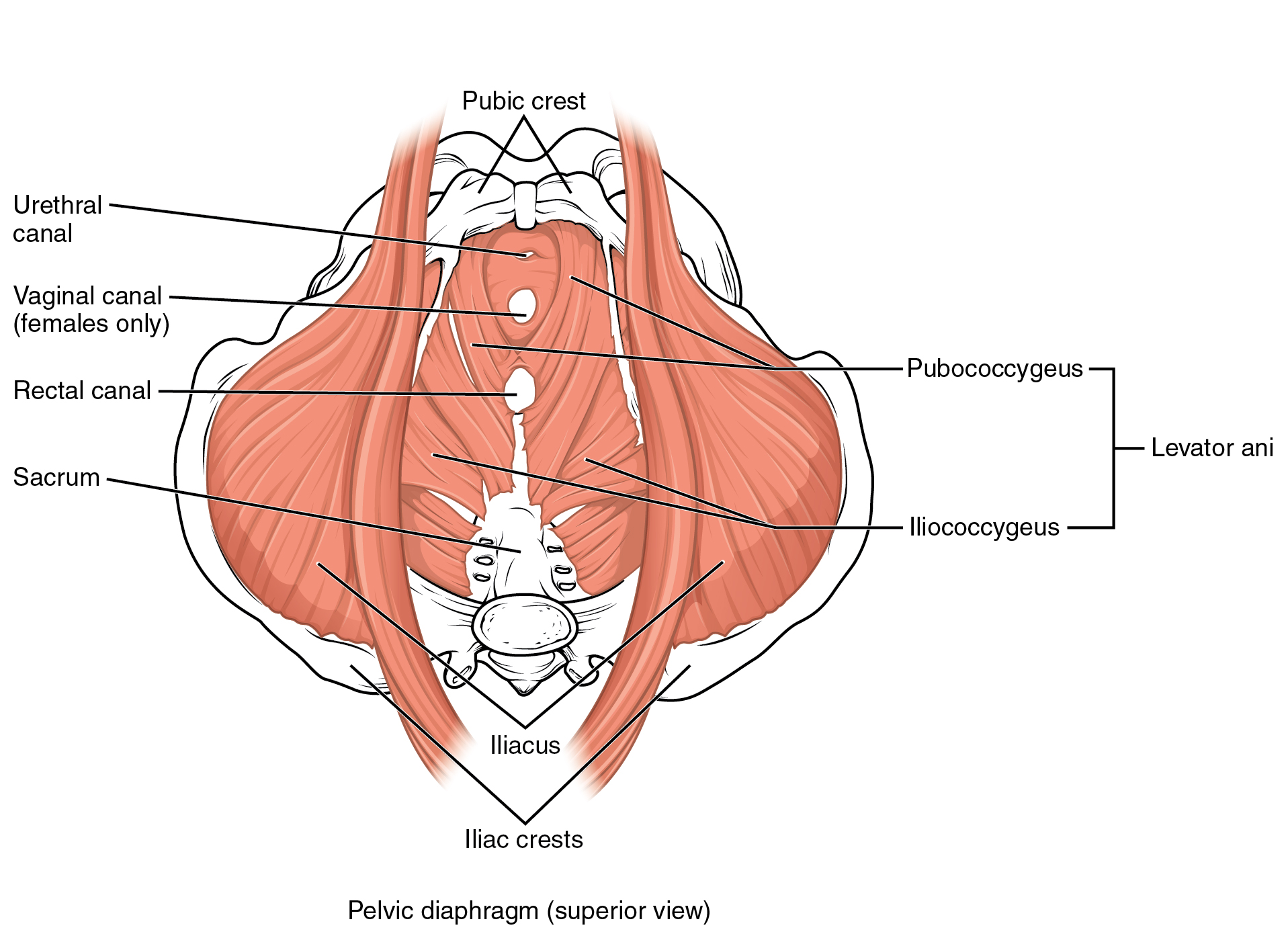
The pelvic floor is a muscular sheet that defines the inferior portion of the pelvic cavity. The pelvic diaphragm, spanning anteriorly to posteriorly from the pubis to the coccyx, comprises the levator ani and the ischiococcygeus. Its openings include the anal culvert and urethra, and the vagina in women.
The large levator ani consists of two skeletal muscles, the pubococcygeus and the iliococcygeus ((Effigy)). The levator ani is considered the about important muscle of the pelvic flooring because it supports the pelvic viscera. It resists the pressure produced past contraction of the intestinal muscles so that the pressure is practical to the colon to aid in defecation and to the uterus to aid in childbirth (assisted by the ischiococcygeus, which pulls the coccyx anteriorly). This musculus too creates skeletal musculus sphincters at the urethra and anus.
Muscles of the Pelvic Flooring
The pelvic flooring muscles support the pelvic organs, resist intra-abdominal pressure, and piece of work as sphincters for the urethra, rectum, and vagina.
The perineum is the diamond-shaped space between the pubic symphysis (anteriorly), the coccyx (posteriorly), and the ischial tuberosities (laterally), lying just inferior to the pelvic diaphragm (levator ani and coccygeus). Divided transversely into triangles, the inductive is the urogenital triangle, which includes the external genitals. The posterior is the anal triangle, which contains the anus ((Figure)). The perineum is also divided into superficial and deep layers with some of the muscles common to men and women ((Figure)). Women also have the compressor urethrae and the sphincter urethrovaginalis, which function to close the vagina. In men, there is the deep transverse perineal muscle that plays a role in ejaculation.
Muscles of the Perineum
The perineum muscles play roles in urination in both sexes, ejaculation in men, and vaginal contraction in women.
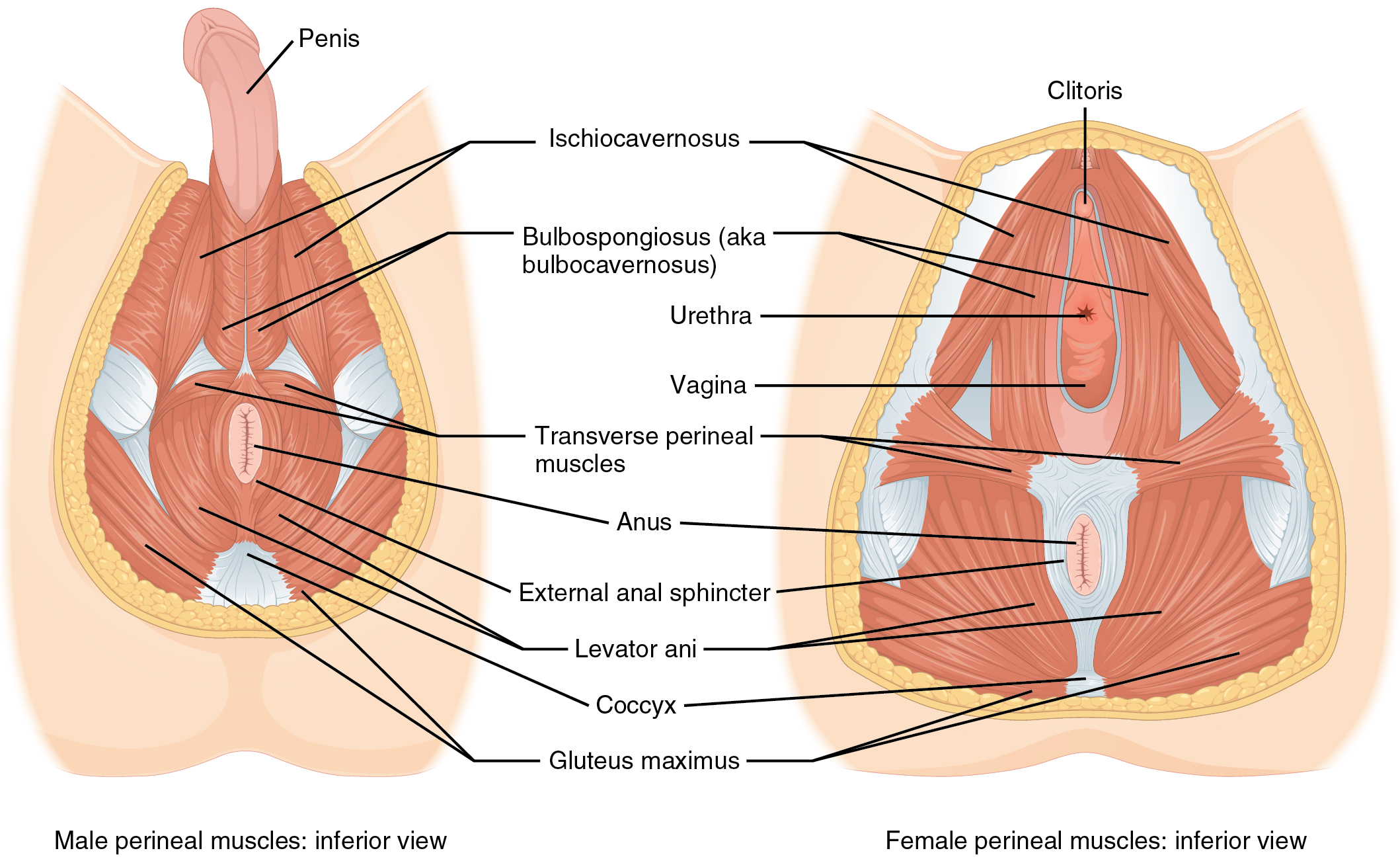
Muscles of the Perineum Mutual to Men and Women
Chapter Review
Fabricated of skin, fascia, and four pairs of musculus, the anterior abdominal wall protects the organs located in the belly and moves the vertebral column. These muscles include the rectus abdominis, which extends through the unabridged length of the trunk, the external oblique, the internal oblique, and the transversus abdominus. The quadratus lumborum forms the posterior abdominal wall.
The muscles of the thorax play a large function in breathing, particularly the dome-shaped diaphragm. When it contracts and flattens, the volume inside the pleural cavities increases, which decreases the pressure within them. Equally a event, air will menstruation into the lungs. The external and internal intercostal muscles span the space between the ribs and help alter the shape of the rib cage and the volume-force per unit area ratio inside the pleural cavities during inspiration and expiration.
The perineum muscles play roles in urination in both sexes, ejaculation in men, and vaginal wrinkle in women. The pelvic floor muscles support the pelvic organs, resist intra-abdominal pressure, and work as sphincters for the urethra, rectum, and vagina.
Review Questions
Which of the following abdominal muscles is not a part of the anterior intestinal wall?
- quadratus lumborum
- rectus abdominis
- interior oblique
- outside oblique
Which musculus pair plays a role in respiration?
- intertransversarii, interspinales
- semispinalis cervicis, semispinalis thoracis
- trapezius, rhomboids
- diaphragm, scalene
What is the linea alba?
- a small musculus that helps with compression of the intestinal organs
- a long tendon that runs down the middle of the rectus abdominis
- a long band of collagen fibers that connects the hip to the knee joint
- some other proper noun for the tendinous inscription
Disquisitional Thinking Questions
Describe the fascicle arrangement in the muscles of the intestinal wall. How practise they relate to each other?
Arranged into layers, the muscles of the abdominal wall are the internal and external obliques, which run on diagonals, the rectus abdominis, which runs directly down the midline of the torso, and the transversus abdominis, which wraps beyond the trunk of the torso.
What are some similarities and differences between the diaphragm and the pelvic diaphragm?
Both diaphragms are thin sheets of skeletal muscle that horizontally span areas of the body. The diaphragm separating the thoracic and abdominal cavities is the principal muscle of animate. The pelvic diaphragm, consisting of two paired muscles, the coccygeus and the levator ani, forms the pelvic floor at the junior end of the trunk.
Glossary
- anal triangle
- posterior triangle of the perineum that includes the anus
- caval opening
- opening in the diaphragm that allows the junior vena cava to pass through; foramen for the vena cava
- compressor urethrae
- deep perineal muscle in women
- deep transverse perineal
- deep perineal muscle in men
- diaphragm
- skeletal muscle that separates the thoracic and abdominal cavities and is dome-shaped at rest
- external intercostal
- superficial intercostal muscles that raise the rib muzzle
- external oblique
- superficial intestinal muscle with fascicles that extend inferiorly and medially
- iliococcygeus
- muscle that makes upwardly the levator ani along with the pubococcygeus
- innermost intercostal
- the deepest intercostal muscles that draw the ribs together
- intercostal muscles
- muscles that span the spaces betwixt the ribs
- internal intercostal
- muscles the intermediate intercostal muscles that draw the ribs together
- internal oblique
- flat, intermediate abdominal muscle with fascicles that run perpendicular to those of the external oblique
- ischiococcygeus
- muscle that assists the levator ani and pulls the coccyx anteriorly
- levator ani
- pelvic muscle that resists intra-abdominal pressure level and supports the pelvic viscera
- linea alba
- white, fibrous band that runs along the midline of the torso
- pelvic diaphragm
- muscular sheet that comprises the levator ani and the ischiococcygeus
- perineum
- diamond-shaped region between the pubic symphysis, coccyx, and ischial tuberosities
- pubococcygeus
- musculus that makes upwardly the levator ani along with the iliococcygeus
- quadratus lumborum
- posterior role of the abdominal wall that helps with posture and stabilization of the body
- rectus abdominis
- long, linear muscle that extends along the middle of the trunk
- rectus sheaths
- tissue that makes up the linea alba
- sphincter urethrovaginalis
- deep perineal muscle in women
- tendinous intersections
- three transverse bands of collagen fibers that divide the rectus abdominis into segments
- transversus abdominis
- deep layer of the belly that has fascicles bundled transversely around the abdomen
- urogenital triangle
- anterior triangle of the perineum that includes the external genitals
Source: https://opentextbc.ca/anatomyandphysiologyopenstax/chapter/axial-muscles-of-the-abdominal-wall-and-thorax/
Post a Comment for "Artlabeling Activity Oblique and Rectus Muscles of the Abdominal Wall Transverse Section"